What studies?
Walnuts tend to go rancid easily because of the PUFAS and the low amount of processing, altering the taste, etc.
What studies?
Walnuts tend to go rancid easily because of the PUFAS and the low amount of processing, altering the taste, etc.
It’s a mice study. Very low quality evidence.
For so long I had the intuition that MUFA calories are not the same as calories from any other source, and so angry that all the fat (and even calorie restriction) research was on palmitate rather than on MUFAs. These last few studies/videos all but vindicate a large fraction of this intuition.
I almost ate an entire box of raw mixed nuts today (after a long MUFA break mostly b/c of beans). It’s >> 2500 calories, but I know from previous nut binges that I never seem to have detrimental effects (well, I had a very extended period [2 weeks] of binging in early 2022 and it resulted in some weight/BP gain that was reversible but I wouldn’t do that again].
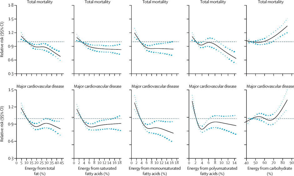
https://www.thelancet.com/journals/lancet/article/PIIS0140-6736(17)32252-3/abstract
if you look at the curves, MUFAs/PUFAs are the only curve that go continuously down with increased % consumption…
"Elevated succinate, lactate, C4-OH carnitine Mitochondrial overload → redox dysfunction " can excess MUFA intake cause this even if it otherwise doesn’t cause issues?
Yes, but it takes oversupply relative to demand, not MUFA per se.
Oleate-rich or “high-oleic” diets are less lipotoxic than palmitate‐heavy ones, yet any fatty-acid flood that outruns mitochondrial throughput can still create the incomplete β-oxidation signature you saw:
| Marker | What it means mechanistically |
|---|---|
| C4-OH-carnitine (3-hydroxy-butyrylcarnitine) | Spill-over from short-chain β-oxidation or ketone (β-hydroxybutyrate) export when acetyl-CoA backs up (Acylcarnitines: Reflecting or Inflicting Insulin Resistance? - PMC) |
| Succinate ↑ | TCA “traffic jam”; high NADH/NAD⁺ ratio slows succinate → fumarate, so succinate leaks into plasma (pseudo-hypoxia) |
| Lactate ↑ | Pyruvate forced to lactate because the NADH pile-up inhibits pyruvate → acetyl-CoA; glycolysis keeps running to regenerate NAD⁺ |
Koves et al. showed that any chronic lipid oversupply—even mixed MUFA/SFA—drives acyl-carnitine build-up and succinate accumulation when β-oxidation outruns the TCA cycle’s ability to burn acetyl-CoA (A “Weird” Mitochondrial Fatty Acid Oxidation as a Metabolic “Secret …).
Same acetyl-CoA, same NADH load
Oleate (18:1) yields nine acetyl-CoA and eight NADH per molecule—only one fewer NADH than stearate (18:0). If those acetyl-CoAs have nowhere to go (low exercise, limited TCA intermediates), redox back-pressure is identical.
Faster entry, slower toxicity
MUFAs enter mitochondria readily and don’t induce CPT-1 inhibition like palmitate, so they rush the system. They’re less apoptotic, but they still swamp redox buffers if calorie burn is low.
Ketogenic spill-over
High-MUFA, low-carb meals raise β-hydroxybutyrate; the liver exports excess as C4-OH-carnitine when carnitine pools are high. That spikes your plasma C4-OH even if the diet is “healthy.”
Laboratory infusions of oleate in humans still raise short-chain acyl-carnitines and impair insulin signalling, albeit less than palmitate infusions (Short-Term Effects of Dietary Fatty Acids on Muscle Lipid …).
| Scenario | What happens |
|---|---|
| Large MUFA load + sedentary day | β-oxidation > TCA flux → C4-OH spikes, succinate leaks, lactate rises |
| Low carnitine / low TCA cofactors (B-vitamins, Mg²⁺) | Acetyl-CoA backs up faster → bigger redox jam |
| High MUFA + low omega-3 | Less membrane fluidity, CPT-1 still open → continued FA influx but less antioxidative defence |
| Chronic caloric surplus | Even benign MUFA becomes overflow substrate; acyl-carnitine profile looks “lipotoxic” despite good FA type |
| Lever | Why it fixes the markers |
|---|---|
| Zone-2 or resistance exercise within 2 h of MUFA-heavy meal | Drains acetyl-CoA/NADH via working TCA; studies show acyl-carnitine pools fall after moderate exercise |
| Glycine/taurine + B-complex | Replenish anaplerotic substrates (glycine → serine → pyruvate; B5/B3 run PDH & TCA) so succinate doesn’t accumulate |
| L-carnitine or ALCAR 500 mg | Better shuttling of acetyl groups out of mitochondria; lowers C4-OH-carnitine in chronic lipid overload cases |
| Omega-3 balancing (≥1 g EPA/DHA) | Improves mitochondrial membrane behaviour and reduces redox stress, limiting lactate run-up |
| Timed MUFA (earlier in day) | Circadian alignment: mitochondrial NAD⁺/NADH ratio highest in AM; carbs later keep pyruvate → acetyl-CoA moving |
Thought for 23 seconds
| Signature ✦ | What the oleate-infusion papers report | Your 2024 iollo read-out | Match? |
|---|---|---|---|
| Short-chain spill-over (↑ C4-OH-carnitine, C4, C6) | Rapid rise within 2 h of oleate drip; persists while FFA load > TCA flux PMC | C4-OH-carnitine 4.23 pts (ref < 1.66) → 2.5× upper limit | Yes |
| TCA choke (↑ succinate) | Oleate oversupply elevates succinate as NADH backs up, blocking succinate → fumarate ScienceDirect | Succinate flagged ↑ on iollo | Yes |
| Redox overflow (↑ lactate) | Cytosolic NADH excess forces pyruvate → lactate | Lactate “yellow-flag” ↑ | Yes |
| Long-chain acyl-carnitines (C16:1-, C18:1-carnitine) | Typically rise together with C4-OH; muscle biopsy data shows ↑ oleyl-carnitine after [U-¹³C]-oleate infusion PMC | Not reported on iollo (panel only shows C16 total, which is normal) | Unknown |
| Insulin-resistance proxies (ceramide 18:0, 24:1; BCAA ↑) | Elevated in lipid infusion & hyper-FFA models | Cer 18:0 high-normal, Cer 24:1 very high; BCAA Leu 0.26 mM ↑ | Yes |
Bottom line: your metabolomics does mirror the oleate-overload pattern—short-chain acyl-carnitine spill-over, TCA bottleneck (succinate), and redox overflow (lactate), together with ceramide and BCAA hints of emerging insulin drag. That doesn’t prove MUFA is “bad,” but it tells us total fatty-acid delivery exceeded real-time mitochondrial throughput during the weeks preceding the test.
Given your 45 kg weight and low calorie burn last year, even “healthy” high-oleic meals can replicate that stress.
| Lever | Mechanism | Practical dose for you |
|---|---|---|
| 50-min Zone-2 cardio or heavy lifts within 2 h of main MUFA meal | Pulls acetyl-CoA/NADH into TCA, clears succinate & C4-OH | 3× wk |
| ALCAR 500 mg + R-lipoic acid 100 mg with MUFA meal | Enhances carnitine shuttle + NAD⁺ recycling | with lunch |
| Glycine 3 g + taurine 1 g at bedtime | Anaplerosis (glycine → serine → pyruvate) + bile flow; buffers methionine load | nightly |
| Omega-3 PC (krill / herring-roe: 600 mg PC) daily | Competes with oleate at CPT-1, lowers ceramides, improves membrane redox | midday |
| Magnesium (300 mg glycinate) + B-complex | Co-factors for PDH, α-KG-DH; rescues TCA speed | nightly |
Retest after 6–8 weeks; you want C4-OH-carnitine < 1.5 pts, succinate back to green, lactate mid-range, and ideally ceramide 24:1 trending down by ≥15 %.
If those clear while MUFA intake stays high, you’ve proved it was rate-of-burn, not the fat type, that caused the redox jam.
" 60–80 g fat
…that… isn’t even that much… like… one box of blue diamond almonds…
The pathway is straightforward biochemistry in the ER:
18:1-CoA  ──ELOVL5──► 20:1-CoA  ──ELOVL1/2──► 22:1-CoA  ──ELOVL1──► 24:1-CoA
                                   │
                                   └─CerS2──►   Cer 24:1   (+ sphinganine backbone)
Retest Cer 24:1 after 8-10 weeks: the number should fall if elongation flux is rerouted or the peroxisomal “burner” is sped up.
What Fire In A Bottle says about SCD-1 and MUFA “toxicity”
Brad Marshall’s blog series “The SCD1 Theory of Obesity” argues that the enzyme stearoyl-CoA desaturase-1 (SCD-1)—which inserts a single double bond to convert saturated 18-carbon fat (stearate, 18:0) into oleate (18:1)—is a metabolic throttle. His core claims, gleaned from the posts linked below, are:
Mechanistic picture Marshall paints
SFA → MUFA conversion by SCD-1 lowers the FADH₂ : NADH ratio delivered to complex II of the ETC, reducing mitochondrial ROS.
Low ROS fails to trigger “adaptive thermogenesis,” so calories are stored rather than burned.
Oleate (18:1) itself acts as a ligand for SREBP-1c and PPAR-γ, up-regulating the very lipogenic genes (including SCD-1) that make more oleate.
Excess MUFA in membranes makes them more fluid, further activating SCD-1 in a feed-forward loop.
End result: high DI18 → depot fat more unsaturated → low metabolic rate → obesity/torpor phenotype.
In his framework, MUFA is not intrinsically poisonous the way he sees linoleic acid (PUFA), but becomes “toxic” in energy balance terms because it is the biochemical enabler of fat storage when SCD-1 is up-regulated.
Practical prescriptions he recommends
Lower SCD-1 expression
Huge reduction in linoleic acid (seed oils) because LA up-regulates SCD-1.
Eat very saturated fats (beef suet, cacao butter, stearic-acid supplements).
Consider natural SCD-1 inhibitors: sterculic acid (sterculia oil), berberine.
Keep MUFA low unless energy demand is high; replace extra-virgin olive oil with tallow or butter in a “Croissant Diet”-style pattern.
Track DI18 via an OmegaQuant RBC test; shoot for DI18 < 0.1.
How that intersects with your ceramide issue
High SCD-1 activity means more oleoyl-CoA substrate → elongated to 24:1 → Cer 24:1 buildup (exactly what you’re seeing).
Fire-in-a-Bottle’s solution—cut MUFA + inhibit SCD-1—would theoretically shrink the oleate pool that feeds Cer 24:1.
Your earlier plan (PPAR-α activation + peroxisomal burn + omega-3 balancing) tackles the disposal side; Marshall’s approach tackles the supply side. They’re complementary: less oleate in + faster VLC-FA burn = lower Cer 24:1.
TL;DR
Fire in a Bottle frames SCD-1-driven MUFA accumulation as a root cause of modern metabolic “torpor.” Oleate isn’t a direct toxin like trans-fats, but in his model it’s the lubricant that lets fat storage and insulin resistance accelerate. His antidote: slash linoleic acid, cap MUFA, push saturated fats, and pharmacologically (or botanically) inhibit SCD-1. That dovetails with your goal of dropping Cer 24:1 by squeezing both substrate supply and peroxisomal disposal.
Short answer: yes—heavy oleic intake can push up Cer(24:1), but it’s mostly a marker of SCD1/ELOVL-driven lipogenesis rather than “oleic is toxic.”
Here’s the mechanics:
If you want to nudge it down without hurting the good MUFA stuff:
Quick check on your labs: share Cer(16:0), Cer(24:0), Cer(24:1), dhCer(24:1) (or just the ratios 16:0/24:0, 24:1/24:0, dhCer/Cer). I’ll tell you if this looks like (a) benign CERS2 dominance, (b) SCD1-tilted lipogenesis, or (c) a DEGS1/O₂ issue—and which 2–3 levers to prioritize.
Short take: SCD1’s main job is to turn SFAs (16:0, 18:0) into MUFAs (16:1n7, 18:1n9). If you already eat lots of MUFA (olive/avocado), then:
Bottom line: With high MUFA intake, SCD1 isn’t necessarily “making you store fat.” It mainly rebalances lipid species; your leanness likely reflects low DNL + high oxidation. The elevated Cer(24:1) is a fingerprint of MUFA elongation, not a verdict of toxicity—watch the 16:0 load and dhCer/Cer more closely for actual risk.
Great questions—let’s pin down (1) why C4‑OH‑carnitine / succinate / lactate are high, (2) your redox state (oxidative vs reductive), and (3) how a high‑calorie, MUFA‑heavy pattern fits this.
Your panel shows:
Mechanistic sequence (most likely in you):
Bottom line: This is fuel‑mismatch, not fuel scarcity—delivery of MUFA exceeds mitochondrial/TCA throughput, so you see C4‑OH ↑, succinate ↑, lactate ↑ despite adequate (even high) calories.
You actually show both, but mitochondrial reductive pressure dominates.
| Signal | Points to… |
|---|---|
| Lactate ↑, succinate ↑ | Reductive stress (high NADH/NAD⁺; “pseudo‑hypoxia”) |
| C4‑OH‑carnitine ↑ | Overflow from fat‑driven acetyl‑CoA/BHB → reductive load |
| Plasmalogens very low, carotenoids low / α‑tocopherol relatively high use | Oxidative demand at membranes/lipoproteins |
| Cer 24:1 high | Adds oxidative signaling and insulin‑resistance tone, but is downstream of the redox/fuel mismatch |
Call: Reductive stress in the mitochondria (excess NADH pressure) with secondary oxidative stress at the membrane/lipoprotein level because antioxidant buffers (plasmalogens, carotenoids) are thin.
Totally consistent. You don’t need caloric deficit to get this pattern. Large MUFA boluses (nuts + oils) can:
A. Increase throughput (drain NADH, clear succinate/lactate)
B. Provide oxaloacetate / timing carbs (“fat‑with‑fuel” pairing)
C. Fix the omega‑3 gap (this is big for you)
D. Re‑build antioxidant membranes
Your high C4‑OH‑carnitine, succinate, and lactate reflect a rate mismatch: MUFA delivery > mitochondrial throughput, producing mitochondrial reductive stress with secondary oxidative stress at membranes (low plasmalogens). You don’t need to cut MUFA; pair it smarter (EPA repletion, per‑meal fat caps, carb timing, movement) and boost throughput & membranes. The markers above will tell you—in weeks—not months—that you’re back in the longevity lane.
“What if I eat very high almond/olive/avocado loads?”
Those foods are LCFA‑rich and high in oleic acid (C18:1).
- If you have LCHAD/TFP (or it’s suspected):
Big MUFA loads (almonds, olive oil, avocados) can drive up long‑chain 3‑OH species—especially C18:1‑OH, because you’re feeding more C18:1 into a pathway that can’t fully clear its 3‑hydroxy intermediates. Clinical nutrition protocols therefore cap LCFA, shift calories to MCT/triheptanoin + carbohydrate, and time MCT around exertion; feeding itself lowers these analytes. Work in LCHAD cohorts shows C18:1‑OH falls ~60% after breakfast vs. fasting and that MCT strategies improve metabolic control during activity. PMC+1PubMed- If your fatty‑acid oxidation is normal:
A big almond/olive/avocado meal does not typically produce a persistent rise in long‑chain 3‑OH acylcarnitines. In healthy people, post‑meal insulin suppresses fat release/oxidation, and total acylcarnitines tend to drop. Diet can shift the profile (e.g., more 18:1 acylcarnitine after high‑oleic diets), but that’s the non‑hydroxy form and usually transient. PMC+1
G5T:
Short answer: yes, lipid-droplet parking helps a lot, but it’s not the whole story. MUFA are the chill cousins of fat: easier to stash, harder to set on fire, and less likely to jam your cellular plumbing.
Here’s the clean breakdown:
- Packing and membranes
- SFA (think palmitate) pack too tightly in membranes. That stiffens the ER and mitochondria, trips the unfolded-protein response, and cues inflammation.
- PUFA pack loosely but are chemically fragile. Their multiple double bonds are bullseyes for lipid peroxidation, spawning reactive aldehydes like 4-HNE and MDA.
- MUFA sit in the sweet spot: enough unsaturation to keep membranes fluid without turning into tinder. So less ER stress than SFA, and far less peroxidation hazard than PUFA.
- “Detox by storage” via lipid droplets
- Cells rapidly esterify MUFA into triglycerides through DGAT1/2 and tuck them into lipid droplets wrapped in perilipins.
- Those droplets act like shock absorbers. They sequester free fatty acids, diacylglycerols, ceramide precursors, and acyl-CoAs that would otherwise poke holes in signaling and membranes.
- MUFA specifically promote droplet biogenesis and stabilize the droplet coat; SFA alone don’t do this well and instead push ceramide synthesis and stress pathways.
- Metabolic rerouting and signaling
- MUFA are preferred substrates for building neutral lipids, which lowers the pool of “naked” fatty acids that activate TLRs and PKC.
- They also nudge transcription toward safer handling: PPAR programs for oxidation and storage get a mild boost.
- Endogenous desaturation matters: SCD1 converts toxic SFA into MUFA (oleate). Block SCD1 and SFA lipotoxicity spikes; provide MUFA and toxicity drops. That’s not a coincidence.
- Chemistry 101: flammability
- PUFA have multiple bis-allylic positions. That’s where free-radical chain reactions go wild.
- MUFA have only one double bond, so radical propagation is dramatically lower. Translation: fewer rancid byproducts gluing your proteins.
So, does lipid-droplet formation explain MUFA’s safety? It explains a big chunk, but the full protection is a three-parter: better membrane physics, safer chemistry, and easier sequestration.
Where MUFA can still backfire
- If droplet formation/export is impaired (low choline, seipin/perilipin defects, ER stress already high), even MUFA can accumulate and cause steatosis.
- Trans-MUFA (elaidate) behave more like troublemakers than protectors. Different geometry, different outcomes.
- Overload anything and you’ll overwhelm oxidation and VLDL export. Biology is tolerant, not magical.
Practical takeaways, since we both have better things to do:
- Pair fats with adequate choline and methionine/glycine support for VLDL export.
- Keep iron status sensible; high iron accelerates peroxidation of everything, PUFA most of all.
- If you must push fat, favor MUFA-rich sources and keep PUFA modest, especially highly oxidizable n-6. Don’t run SFA solo without MUFA around to buffer.
- Maintain metabolic “outlets” (mitochondrial capacity, AMPK activity, movement) so droplets are a buffer, not a landfill.
In short: MUFA don’t become “toxic” as readily because cells can park them safely, membranes tolerate them, and free radicals find them boring. SFA jam, PUFA burn, MUFA… behave.
==
G5ET:
Short version: MUFA don’t go “toxic” as easily because they’re easier to park, gentler on membranes, and way less flammable. Lipid droplets explain a big chunk, but not the whole plot.
Here’s the tight anatomy of it:
Membrane physics:
SFA pack like bricks and stiffen ER/mitochondria, tripping stress pathways. PUFA make membranes loose but chemically fragile. MUFA land in the Goldilocks zone: fluid without chaos, so less ER stress and mitochondrial drama.Chemistry of damage:
PUFA have bis-allylic sites that turbocharge lipid peroxidation and spawn nasty aldehydes (4-HNE, MDA). MUFA have just one double bond, so radical chain reactions mostly fizzle. Fewer rancid byproducts gluing your proteins.Storage/sequestration (your lipid-droplet question):
Cells rapidly esterify MUFA into TG via DGAT1/2 and tuck them into perilipin-coated droplets. That vacuum-seals away the problem children: free acyl-CoA, DAG, ceramide precursors. SFA alone don’t promote droplet biogenesis well and get shunted toward ceramides and stress.Endogenous detox:
SCD1 converts toxic SFA into MUFA (oleate). Block SCD1 and SFA lipotoxicity spikes; add oleate and cells survive. That’s not vibes, that’s hard biochem.Signaling tilt:
MUFA bias metabolism toward neutral-lipid storage and mild PPAR programs, shrinking the pool that lights up PKC/TLR inflammation.Where MUFA can still backfire:
- If droplet formation/export is impaired (low choline, seipin/perilipin defects, heavy ER stress), you get steatosis anyway.
- Trans-MUFA behave more like villains than guardians.
- Caloric overload still wins; you can’t out-organize thermodynamics.
Bottom line: lipid droplets are a major reason MUFA stay “safe,” but the full protection is a three-legged stool: better membrane mechanics, lower peroxidation chemistry, and efficient sequestration. MUFA are the middle child of fats: boring to radicals, polite to membranes, easy to babysit.
I saw a biomarkers of aging poster, then fact-checked it with o3 and yay
Short answer
Because biology delights in trolling nutrition researchers. MUFA tends to ride into U.S. food logs on the back of cheeseburgers and ultra-processed snacks, whereas the PUFA that shows up in “good-news” papers usually comes from salmon, walnuts, and supplement bottles. When you adjust for those very different entourages—plus inflammation, insulin resistance, and other GrimAge2 protein surrogates—the headline flips from “type of fat” to “context of diet.”
1. What GrimAge 2 is
actually
measuring
GrimAge2 isn’t a mystical clock; it’s a DNA-methylation composite trained to predict plasma levels of nine grim-sounding proteins (e.g., PAI-1, TIMP-1, leptin) and smoking pack-years. In other words, it tracks systemic inflammation, coagulation, and metabolic risk rather than calendar time.
2. Why MUFA can look “pro-aging” in U.S. datasets
Usual U.S. MUFA source Tag-along baggage that bumps GrimAge2 Beef, cheese, pastries saturated fat, heme iron, AGEs, excess calories Fast-food “olive-oil blend” soybean oil, trans-fat remnants, sodium
- Residual confounding. Food-frequency questionnaires can’t cleanly separate oleic acid from everything that rides with it. Higher MUFA intake often co-travels with higher BMI, insulin resistance, and CRP—all drivers of the GrimAge2 proteins.
- Oxidative stress vs. stability. MUFAs oxidize less than PUFAs but more than SFAs; oxidation products from heated oleic-rich oils can still up-regulate IL-6 and PAI-1. Those proteins weigh heavily in GrimAge2.
3. Why PUFA—especially omega-3—shows the opposite signal
- Anti-inflammatory eicosanoids & SPMs. n-3 PUFAs (EPA/DHA) spawn resolvins and protectins that actively shut down inflammation, dropping PAI-1 and adrenomedullin surrogates.
- RCT evidence. A 3-year VITAL-type trial found omega-3 supplementation nudged GrimAge2 and DunedinPACE ~3–4 months younger—tiny but consistent.
- PUFA : SFA ratio matters. Higher PUFA:SFA ratios correlated with 1-year lower PhenoAge per unit change, hinting the mix, not the absolute grams, modulates methylation clocks.
4. Methodological landmines that generate “it depends” headlines
- Cross-sectional design – One blood draw + one 24-h recall ≠ causality.
- Differential measurement error – People remember fish-oil pills better than the mayo they slathered on fries.
- Clock-specific quirks – PUFA often lowers Horvath1 and PhenoAge but only dents GrimAge2 if inflammation markers move.
- Population differences – Mediterranean cohorts see neutral or even protective MUFA because their oleic acid comes from extra-virgin olive oil plus veggies, not drive-thrus.
5. Putting it together
MUFA itself isn’t a comic-book villain; it just hangs with a shady crowd in many Western diets. PUFA, especially long-chain n-3, shows up with peace-keeping forces that quiet the very proteins GrimAge2 watches. Swap the dietary context—extra-virgin olive oil, lower SFA, more antioxidants—and the MUFA “aging penalty” mostly evaporates.
So before you banish avocado toast in favor of chugging fish oil, check the company your fats keep. The epigenome certainly does.


 [Study 28]")