What is your RNFL thickness (retinal nerve fiber layer thickness)? Easily-measured indicator of brain/inner-cell health

https://iovs.arvojournals.org/article.aspx?articleid=2127832

Table 1

Age, SE, axial length and 360° RNFL thickness in EM, LM, MM and HM groups

Variables EM LM MM HM
Age (y) 31.85±10.55 31.10±9.64 31.38±11.03 31.15±8.68
SE (D) -0.11±0.21 -1.47±0.73 -4.45±0.81 -8.25±1.41
Axial length (mm) 23.18±0.75 23.74±0.70 25.03±0.85 26.37±1.35
Average RNFL thickness (µm) 100.91±10.07 98.68±8.81 94.97±9.02 89.88±9.41

=> this is why it’s important

There was a statistically significant difference in the average RNFL thickness between the three degrees of myopia groups, with least myopia having higher RNFL thickness and high myopia with least RNFL thickness (P = 0.001); low myopia group mean RNFL being 92.17 (+9.84), moderate myopia group 88.12 (+9.53) and high myopia group 82.40 (+10.

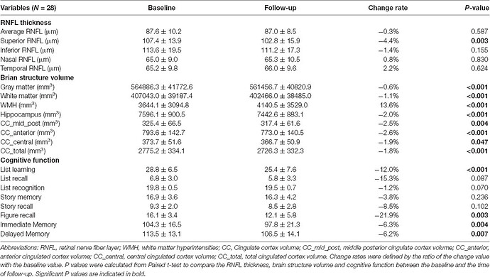
Recently, two population-based cohort studies further reported that thinner RNFL was associated with current and future cognitive impairment, and could predict the incidence of dementia within 4.5 years in nondemented participants (Ko et al., 2018; Mutlu et al., 2018). These findings suggested that retinal and brain neurodegeneration may occur in parallel to some extent, and RNFL thickness could serve as a noninvasive and less expensive biomarker of AD.

Given the wide variance of RNFL thickness in old adults, OCT measurement at a single time point may not be informative enough to the identification of abnormal RNFL profiles in AD patients (Mok et al., 2002). In our previous studies, we found that longitudinal reduction of RNFL thickness was associated with the decline of episodic memory in old adults (Shen et al., 2013), and those who suffered cognitive decline within 25 months had more reduction of RNFL thickness with respect to old adults with stable cognition (Shi et al., 2014, 2016). It supports the hypothesis that a longitudinal change of RNFL thickness would be more indicative of cognitive deterioration among the elderly population
2 Likes

Thinning rates were classified according to SE into three groups: nonmyopic (NM; >0 D), mild-to-moderately myopic (MM; >–6 D and ≤0 D), and highly myopic (HM; ≤–6 D). Results. The overall slopes of change in RNFL thickness over time in the NM, MM, and HM groups were −0.305 ± 0.128, −0.294 ± 0.068, and −0.208 ± 0.097 μm/yr, respectively. Slopes of RNFL thickness changes in these groups were −0.514 ± 0.248, −0.520 ± 0.133, and −0.528 ± 0.188 μm/yr, in the superior quadrant; −0.084 ± 0.145, 0.107 ± 0.082, and −0.161 ± 0.112 μm/yr, in the temporal quadrant; −0.807 ± 0.242, −0.794 ± 0.130, and −0.727 ± 0.183 μm/yr, in the inferior quadrant; and 0.160 ± 0.157, 0.118 ± 0.084, and 0.429 ± 0.119 μm/yr, in the nasal quadrant. Overall and in all four quadrants, there was no significant difference in the rate of RNFL thickness change among the three groups. Conclusions. Refractive error did not affect the physiologic thinning rate of RNFL when assessed by SD OCT.

Similarly, thickening of the RNFL in this quadrant by +0.308 μm/yr has been reported [2], further suggesting that the nasal sector did not experience age-related loss in RNFL thickness. This may have been due to the proportion of nonneuronal tissue, such as glial tissue, in the RNFL, which has been reported to increase with age [27, 28]. OCT assesses the thickness between the internal limiting membrane and the ganglion cell layer in the retina; thus, OCT cannot measure the RNFL separately from other layers. Axonal fibers in the RNFL decrease with age, indicating an inverse relationship between thickness and the proportion of nonneuronal tissue. Thus, changes in RNFL, as measured by OCT, result from a combination of a decreased width of neuronal tissue and an increased width of nonneuronal tissue. This would apply not only to the nasal quadrant but also to all quadrants. However, consistent results showing that RNFL thickness in the nasal sector increases or remains stable suggest an effect of nonneuronal tissue and its possible increase over time.

In contrast to our results, RNFL thinning rate has been reported to be faster in highly myopic than in nonmyopic eyes in subjects aged 40–59 years who were followed-up for more than 3 years [29]. Because our subjects were relatively younger (mean age, 40 ± 12 years), direct comparison between these studies may be inappropriate. Furthermore, the previous study also reported that RNFL thinning rates were similar in highly myopic and nonmyopic eyes of younger subjects, in agreement with our results [29]. Furthermore, the shorter follow-up period in the previous study (3 years) than in ours (6 years) may have resulted in different outcomes. Highly myopic eyes with injured and thinner RNFL in younger subjects due to a sudden increase in axial length may experience a faster deterioration of the RNFL as the subject becomes 40–59 years old. Further studies are needed, however, to test this hypothesis. Our participants showed faster thinning in men compared with women. This may be due to thinner baseline RNFL in men than in women (83.4 vs. 87.7 μm).

Although the exact pathophysiology of RNFL thinning in CVD remains unknown, it has been postulated to be due to the impairment of ocular circulation. The retina is supplied by the dual end arterioles, central retinal artery, and short posterior ciliary arteries, with autoregulatory mechanisms to maintain ocular perfusion. In the case of subclinical cardiovascular and cerebrovascular abnormalities, retinal vasoconstriction reduces blood supply to the retina in favor of adequate perfusion to the systemic circulation and important organs, leaving the inner retinal layers susceptible to ischemic damage [7]. It is therefore probable that microvascular pathologies, including atherosclerosis, arterial hypertension, increased rigidity and insufficient autoregulation, could be the main driver behind the reduction of RNFL thickness [46]. Moreover, retinal ischemia/reperfusion induced oxidative stress injury, cytokines release, and nerve fiber death might contribute to secondary RNFL thinning [47]. However, further studies are warranted to uncover the relevant underlying mechanisms.

The most notable advancement in ophthalmology was the advent of high-resolution OCT technology, a rapid, non-invasive and widely available imaging modality capable of producing a high-resolution cross-sectional image reflecting the near-histological tissue microstructure in vivo [48, 49]. As the only human tissue that allows direct non-invasive visualization of microvascular circulation and the central nervous system, the retina provides a unique window for documenting systemic diseases [48, 50, 51]. OCT examination is increasingly being routinely performed in hospital and community settings. The number of OCT scans increased 14-fold from 23,500 scans in 2008 to over 330,000 scans in 2016 at Moorfields Eye Hospital NHS Foundation Trust [52]. Furthermore, it has been reported that healthcare-seeking behavior for eye health has surpassed that for cardiovascular disease [49, 53], which provides OCT with an unprecedented opportunity to detect systemic disease, predict its onset, and quantify its severity and response to treatment. With the emergence of automated segmentation and precise quantification of individual retinal layers by deep learning algorithms [54], our discovery provides a novel insight into the role of OCT derived morphological RNFL abnormalities in the pathologies of cardiovascular events.

1 Like

The global RNFL thickness decreased with age by 4.97 μm per decade (β = -0.497; p = 0.021), and the segmental RNFL thickness significantly decreased in the superonasal (-9.90 μm per decade, p = 0.001) and temporolower (-6.78 μm per decade, p = 0.001) segments; the same trend showed borderline significance in the

1 Like

For Bryan Johnson:

Eye Health

MEASUREMENT PROTOCOL

Drusen volume 0
Enhanced depth imaging optical coherence tomography subfoveal choroidal thickness high n=1 accuracy read left age 51, right age 70, prior to axial length/refractive error regression adjustment
IOP 13 mmHg, age 38
Accommodative distance high n=1 accuracy read TBC
Eyelash length age 70+ (genetically short) 

EYE MEASUREMENTS

Sub-foveal choroidal thickness on enhanced depth imaging spectral domain optical coherence tomography (sfCT EDI-SD-OCT) was acquired showing 332 microns and 400 microns respectively; however, future 3D choroidal quantification methods we will perform will have lower standard deviation and higher biological age resolution