Parkinson's disease

@John_Hemming’s theory vindicated? Just published: Citric acid disassembles α-synuclein fibrils and reduces their cytotoxicity 2025

:warning: Korean paper from an okay but not Tier 1 institution + Short publication and not long paper + Mechanistic in vivo model :warning:

Misfolding and aggregation of α-synuclein are associated with the progression of Synucleinopathies including Parkinson’s disease
Citric acid can disrupt the β-sheet structure of α-synuclein fibrils and inhibit additional fibril formation
The molecular docking simulation shows that citric acid exhibits a strong affinity for β-sheet stacking and adjacent regions
Citric acid reduces the cytotoxicity of α-synuclein fibrils

image

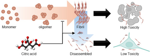
α-synuclein plays a crucial role in regulating neurotransmitter release and synaptic plasticity [1]. However, the misfolding and aggregation of α-synuclein are major hallmarks of Lewy bodies related to progressive neurodegenerative disorders, including Parkinson’s disease, dementia with Lewy bodies, and multiple system atrophy [2]. Thus, developing therapeutics that target α-synuclein aggregates is crucial for treating synucleinopathies, as it alleviates neurotoxicity [3]. Herein, we demonstrate that citric acid (citrate, 2-hydroxy-propane-1,2,3-tricarboxylic acid), commonly found in food supplements, can disassemble α-synuclein fibrils and mitigate their neuronal toxicity (Fig. 1A). Our findings reveal that citric acid can effectively disrupt β-sheet structure of α-synuclein fibrils and inhibit further fibril formation. Molecular docking (MD) simulation further supports these results, showing that citric acid exhibits a high affinity for the β-strand and the surrounding region within α-synuclein fibrils. We propose that citric acid holds significant promise for pharmaceutical applications in treating neurodegenerative diseases related to synucleinopathies.

In summary, we demonstrated that citric acid can effectively inhibit the α-synuclein fibril formation and disaggregates existing fibrillar aggregates. It also reduces the cytotoxicity of α-synuclein fibrils and interacts with the β-strand regions and their surrounding residues within the fibril structure. Given that pathological α-synuclein aggregates may originate in the gut, orally administered citric acid could act locally to disassemble them before brain propagation. Notably, although typical plasma citrate levels are around 0.1 mM, gastrointestinal concentrations can transiently reach 10–20 mM after citrus ingestion, supporting the feasibility of our effective dose for gut-targeted intervention.

1 Like

My theory is moreso about acetylation.

This, however, would help particularly with PD

3 Likes

Yes. I find the closing remark about gut concentrations interesting. Worth exploring…

1 Like

Obviously the gut will be relatively high, but serum is unlikely to get over 0.5mM

1 Like

It’s well known that coffee is associated with a lowest risk of PD. However, RCT failed. A new Mendelian randomization clarifies the situation: Coffee Consumption Is Associated With Later Age-at-Onset of Parkinson’s Disease 2025

Using Mendelian randomization, we identified a significant association between coffee consumption and delayed PD AAO (IVW: OR, 1.91; 95% CI 1.53–2.38; p = 8.072e-09), but no causal association or genetic correlation with PD risk or progression. Our findings suggest a potential causal effect of higher coffee consumption on PD AAO, with no evidence of an association with PD risk or progression.

On another topic and relevant to those taking immunosuppressants such as rapamycin? Prognostic significance of peripheral lymphocyte counts in Parkinson’s disease 2025

PD patients who reached an endpoint had lower lymphocyte counts at baseline.
PD patients with low lymphocyte counts reached an endpoint earlier.
Motor symptoms and lymphocyte count were associated with reaching an endpoint.

They don’t define the threshold between the “low lymphocyte” and “high lymphocyte” groups but they give the groups’ averages (×10^3/μL): 1.10±0.25 (low) vs 1.87±0.44 (high).

Do you have any thoughts on this @John_Hemming?

WBC issues are complicated.

In theory a low aggregate WBC count is pro longevity, but within that a high proportion of lymphocytes is better.

Hence simply looking at the lymphocyte count ignores the conflicting points.

1 Like

Speculation for those with an extra few minutes at lunch. FWIW:

What’s Causing the Parkinson’s Belt?

I posted a study before about the geographic, sex, and ethnic prevalence of PD in the US. The topic is a little noisy.

1 Like

Same author (Tiago F. Outeiro) just published: Circadian clock dysfunction in Parkinson’s disease: mechanisms, consequences, and therapeutic strategy 2025

In addition, atrophy of the SCN previously observed in histology experiments43, which may explain the weakened body temperature and altered melatonin rhythms under the direct SCN control. Altogether, these underscore neurodegeneration in specific brain regions may contribute to the circadian alterations observed in PD patients. However, the precise role of circadian dysfunction in the onset and progression of these symptoms remains further to be elucidated.
Circadian disruptions in PD significantly impact hormonal secretion, including cortisol and melatonin, as well as core body temperature and blood pressure rhythms, all of which are directly regulated by the circadian clock53,54. In a study involving 30 PD patients and 15 controls, plasma cortisol levels were found to be elevated in PD patients, whereas circulating melatonin was reduced despite no observable change in the phase of the rhythms55. A recent systematic review investigating cortisol alterations in PD identified out of 21 studies assessed ten studies reported disrupted cortisol rhythms, with the majority (seven out of ten) indicating elevated cortisol levels56. However, some studies failed to find significant changes or even observed opposite trends, highlighting the need for further research to validate these findings. Conversely, plasma melatonin levels have been shown to be significantly lower in PD patients compared to controls, as measured by radioimmunoassay in a study with 20 PD patients and 15 controls57. Another study, which included PD patients undergoing medical treatment (N = 16), unmedicated patients (N = 13), and healthy controls (N = 27), revealed that medicated patients exhibited a longer phase angle of entrainment (see Box 2 for definition) indicating a larger time difference between phase of the circadian rhythm and the external cue, calculated by subtracting salivary dim light melatonin onset (DLMO) from habitual sleep onset58. In addition, higher melatonin levels were observed compared to the non-medicated group58. These findings suggest that alterations in melatonin rhythms could be influenced by dopaminergic treatments (Box 1).
In humans, melatonin is one of the best studied compounds in the context of restoring circadian dynamics, which has been shown to enhance subjective sleep quality in PD and to exhibit antioxidative properties104,105. A slight improvement in nocturnal sleep in PD patients taking 50 mg of melatonin (in comparison to 5 mg placebo) has been observed, though the improvement was short-term (roughly 10 min)106. Medeiros and colleagues on the other hand observed improved subjective sleep quality in PD patients taking a much lower dose (3 mg/day) of melatonin for a month, despite no significant change was detected in polysomnography (PSG) results107. Delgado-Lara et al. reported increased BMAL1 gene expression in PD patients who were administered 25 mg of melatonin for 3 months, particularly in the morning, suggesting the improvement in PD symptom management is linked to restoration of core-clock machinery. In a recent systematic review where seven randomized controlled trials were assessed, melatonin was suggested as a safe and well-tolerated compound for the management of insomnia in PD patients albeit there was no improvement in daytime sleepiness or RBD symptoms109. Other evidence showed a positive improvement of motor and nonmotor PD symptoms (including sleep disorders) in PD patients following treatment with cannabis110. With the development of new pharmacological compounds, such as small-molecule modulators, a restoration of disrupted CR has been explored. For example, compounds that inhibit casein kinases led to period-lengthening effects in human osteosarcoma cells (U2OS) and mouse embryonic fibroblasts (MEFs)111, proposed to be linked to neuroprotective actions of CKI-δ encoded by CSNK1D. CKI directly acts on core-clock post-translational modifications by phosphorylation of PER23 pending to be validated further in vivo. Despite these exciting developments, the exact molecular mechanisms behind clock disruption and PD are yet to be further elucidated. Moreover, clinical studies that take individual circadian rhythms into account are scarce, and further research in this field is timely.
Furthermore, interventions to revert circadian changes, including BLT or melatonin supplements, have shown promising benefits in improving both motor and nonmotor symptoms.

Would vasopressin or the VIP peptide be of any help in restoring SCN function or enhancing it or replacing it?

"Astounding” Results: Blocking One Enzyme Brings Parkinson’s-Damaged Cells Back to Life

https://scitechdaily.com/astounding-results-blocking-one-enzyme-brings-parkinsons-damaged-cells-back-to-life/

Interesting!

1 Like

Exciting! As noted in the article this is primarily relevant to people with genetic PD (less than 10% of all cases) but “The next step for the research team is to test whether other forms of Parkinson’s disease that are not associated with the LRRK2 genetic mutation could benefit from this type of treatment.” Wait and see…

1 Like

On calcium and PD @CronosTempi (@DrFraser you might also be interested):

Many papers previously found a protective association between calcium-channel blockers (CCB) use and PD.

However, the RCT of isradipine (a brain-penetrant CCB) failed: Isradipine Versus Placebo in Early Parkinson Disease A Randomized Trial 2020

There’s still an ongoing trial of nilvadipine (4 mg twice a day) in Australia, results expected soon.

But a recent Yale preprint shows a different picture: Drug Repurposing for Parkinson’s Disease: A Large-Scale Multi-Cohort Study 2025

They did find a lower risk of PD among amlodipine users: "Two commonly used antihypertensives, amlodipine and losartan, were also linked to a lower PD risk.”

But after diagnosis, they found a faster decline among amlodipine users: “After adjusting for covariates, PD patients who had ever used sildenafil or amlodipine exhibited a significantly faster cognitive decline in MoCA scores (sildenafil: β = –0.13, p = 0.019; amlodipine: β = –0.11, p = 0.027) and a significantly faster motor decline by UPDRS III scores (sildenafil: β = 0.83, p = 0.000073; amlodipine: β = 0.66, p = 0.0032), compared to those who had never used these drugs (appendix p 34).”

So calcium channel blockers protect you before getting PD, but once you get PD, they make you worse off? When does PD even start?

A recent paper sheds a new light on that conundrum: The molecular clock drives motivated locomotion and time-of-day-dependent firing patterns in mouse dopaminergic neurons 2025

They found that the bursting of nigral dopamine neurons in mice is blocked by the L-Type CCB N-nifedipine. According to the paper, this bursting is time-of-day dependent, Bmal1-regulated, and reliant on L-type Ca²⁺ channels (LTCC) during early night periods. This is Grok’s interpretation of the paper:

On one hand, the paper highlights a potential beneficial aspect: Aging increases reliance on LTCCs for pacemaking in substantia nigra pars compacta (SNc) DANs, which elevates cellular stress and has been implicated in the heightened vulnerability of these neurons in PD. This suggests that reducing excessive LTCC activity—possibly through blockers—could mitigate stress and offer neuroprotective effects, aligning with the paper’s discussion of how circadian clock disruptions (e.g., via Bmal1 deletion) might exacerbate metabolic demands and neurodegeneration in PD-relevant pathways.
On the other hand, the study shows a potential detrimental effect: LTCCs are essential for time-of-day-dependent bursting in SNc DANs during the early night (ZT 12–16), which is Bmal1-regulated and critical for phasic dopamine release and motivated locomotion. Experiments using the LTCC blocker nifedipine completely abolished this bursting without affecting tonic firing, implying that acute blockade could impair normal physiological outputs, such as voluntary movement initiation, which are already compromised in PD.

What does this mean for humans? According to Grok again, mice are nocturnal, while humans are diurnal. So you want to block LTCC during the night (to protect the brain) but not during the morning/ early day (to allow for daytime bursts and support motivation and movement).

I noticed this myself: when I’m on amlodipine (a long-acting CCB) I’m better overall but a bit slower and more apathetic. When I’m off amlodipine, I’m more energetic and faster, but I have slight tremors.

So is there a solution? If the paper is correct and applicable to humans, then you want an L-type CCB with a short half-life that you can take once daily in the evening to protect the brain at night while keeping LTTC active during the day. Ideally this CCB should be brain-penetrant. And this brings us back to… isradipine! Indeed: “The half-life of isradipine is biphasic, with an alpha half-life of 1.5 to 2 hours and a terminal half-life of approximately 8 hours.”

So why did the isradipine trial fail then? I think because they used “5 mg of immediate-release isradipine twice daily or placebo for 36 months.” My guess is that the evening dose was beneficial, while the morning dose was detrimental, resulting in a net effect of 0. Would they find benefits with isradipine IR, taken once daily in the evening?

I’d like to try isradipine IR, but it appears to be available only in the US and Canada. I couldn’t find it on IndiaMart or in Turkey. Some countries have isradipine slow-release but this defeats the purpose.

@John_Hemming: Magnesium being a very mild L-type CCB, this might explain why it doesn’t have significant benefits in PD?

3 Likes

Has this been posted? I did a search but couldn’t find it. Very old paper.

Use of Calcium Channel Blockers and Parkinson’s Disease

Not posted but that’s the paper I had in mind when I wrote “Many papers previously found a protective association between calcium-channel blockers (CCB) use and PD.” Others replicated the finding.

1 Like

As usual, a great post!

I think the delay of onset of PD is probably the most relevant item for many individuals - and different approaches may be needed once one has disease, and may need to change with changing severity.

I currently think that the neuroprotective approaches for AD and PD are likely quite similar pre-disease, but need to start 20 years before one would have had a diagnosis of AD or PD.

The big issue is the need to change strategies and when those change, and on what evidence when dealing with early PD, advanced PD, or MCI, then early AD.

Once disease is present, especially once able to be diagnosed as having PD or AD it is more difficult to substantiate what modifies disease/progression. The sluggish movement of studies and data mining is going too slowly for those who have early disease, so many have to take their best guess based on inadequate information.

We know a lot more about how to risk decrease for AD, there is less on PD - however, many of the same interventions show significant risk reduction, such as Mediterranean diet, exercise, vitamin E, statins, CCBs, NSAIDS, baseline cognitive function, and others.

On the CCB question - you may look at these options - especially the first one as it has high penetration to brain:
|Drug|Half-life|BBB Penetration|
|Nimodipine|~1–2 h|High|
|Nicardipine|~2–4 h|Moderate|

1 Like

I was looking at nifedipine as a short acting, and saw that you already posted the paper showing amlodipine with better ACM compared to nifedipine or felo, vera etc.

Calcium channel blockers, survival and ischaemic stroke in patients with dementia: a Swedish registry study

The problem is by the time I come across a paper, more likely than not, you already posted it :joy:, like below

Brain-penetrant calcium channel blockers are associated with a reduced incidence of neuropsychiatric disorders

I’m keeping my eye out on calcium handling, as I sense that this is the most promising practical approach to ameliorating early PD.

2 Likes

As usual, I agree with you :wink:

Nimodipine needs to be taken on an empty stomach (≥ 1 h before or ≥ 2 h after food) and can cause tachycardia. Nicardipine appears promising, but in many countries, it’s only available in IV form. I’ll have to do more research…

1 Like

Nicardapine 20 mg 90 capsules in U.S. with a GoodRx coupon is not that cheap - but $178 at CVS. ?if India has these?

1 Like

Metabolic Dysregulation in Parkinson’s Disease: Non-Oxidative Phosphorylation and Its Role in Brain Energy Metabolism 2025

Parkinson’s disease (PD) is a progressive neurodegenerative condition affecting around 1-2% of the population over the age of 60. The lack of disease-modifying therapies highlights the need for insights into the etiology and pathogenesis of PD. Mitochondrial dysfunction is recognized to be a significant contributor to disease pathogenesis, resulting in bioenergetic deficits and subsequent neurodegeneration. Research indicates that changes in non-oxidative phosphorylation (non-OXPHOS) metabolism in PD may serve as an adaptive response to mitochondrial dysfunction, compensating for energetic failure and alleviating disease progression. This review explores mitochondrial dysfunction-driven alterations in non-OXPHOS metabolic pathways, such as glycolysis and the tricarboxylic acid cycle, emphasizing their role in maintaining energy metabolism and their dual contribution to neuroprotection and disease progression. Advances in neuroimaging techniques are also discussed, particularly their role in visualizing metabolic changes in vivo and their potential utility in identifying non-OXPHOS metabolism as a biomarker of mitochondrial dysfunction. By enhancing our understanding of the complex interplay between metabolic pathways in PD, this review underscores the importance of personalized therapeutic approaches that consider individual metabolic variations. Ultimately, these insights aim to pave the way for improved diagnostic utility and personalized treatment strategies that address the metabolic and mitochondrial dysfunctions underlying PD pathogenesis.

Full paper: PDF

@John_Hemming: non-OXPHOS and TCA cycle :thinking:

On glycolysis: “The evidence of glycolysis’ role in PD is conflicting, demonstrating the upregulation of specific glycolytic enzymes as a neuroprotective mechanism, while increased glycolytic flux results in the accumulation of toxic end-products, exacerbating neurodegeneration. The dual nature of glycolysis necessitates identifying the threshold balancing the compensatory benefits against neurotoxic effects. The lack of consistency suggests that the glycolytic reprogramming in PD is context-dependent, with factors such as cell type, disease stage, underlying genotype, and threshold of glycolytic flux shaping its contribution to disease progression.”

On TCA cycle: "This process is significantly disrupted in PD, as evidenced by findings from in vivo PD models. PDH catalyzes the first gatekeeping step of the TCA; however, its activity is compromised in PD. […] The consistent evidence of reduced PDH activity suggests TCA cycle decoupling from glycolysis, leading to elevated pyruvate levels and reduced glucose flux through oxidative metabolism. Elevated pyruvate levels are converted to lactate due to increased LDH activity in PD, signifying a metabolic shift towards anaerobic glycolysis. […] MPP±treated SH-SY5Y cells demonstrated reduced citrate synthase (CS) and isocitrate dehydrogenase activity (IDH)

Fatty acid β-oxidation: “Similarly to glycolysis, FAO appears to be differentially affected across disease stages – upregulated in early stages to meet energy demands and declining with the disease progression; however, this pattern remains controversial and not fully supported by all research available. […] Evidence linking dysregulated FAO to pathogenesis is increasing; for instance, protein analysis of post-mortem tissue revealed early-stage FAO upregulation, marked by increased expression of acetoacetyl-CoA thiolase, a key FAO enzyme [37]. Reduced mitochondrial complex I activity correlated with abnormal FAO, marked by increased plasma levels of isobutyrylcarnitine, reinforcing FAO dysregulation as a consequence of mitochondrial dysfunction [38]. Despite the evidence from some studies reporting FAO upregulation, its overall function is disrupted, as indicated by a shift from FA metabolism to lipid storage in PD.”

I need to read this when I have some time, but it all seems to report the consequences of a failing neuron being downstream of damage to Complexes 1 and 3.