Very complex stuff.
Worth considering a calcium channel blocker for hypertension and autophagy thrown in the mix.
Very complex stuff.
Worth considering a calcium channel blocker for hypertension and autophagy thrown in the mix.
Another thought is combination of rapamycin with lithium for additional autophagy.
ABSTRACT
We recently showed that lithium induces autophagy via inositol monophosphatase (IMPase) inhibition, leading to free inositol depletion and reduced myo-inositol-1,4, 5-triphosphate (IP3) levels. This represents a novel way of regulating mammalian autophagy, independent of the mammalian target of rapamycin (mTOR). Induction of autophagy by lithium led to enhanced clearance of autophagy substrates, like mutant huntingtin fragments and mutant α-synucleins, associated with Huntington’s disease (HD) and some autosomal dominant forms of Parkinson’s disease (PD), respectively. Similar effects were observed with a specific IMPase inhibitor and mood-stabilizing drugs that decrease inositol levels. This may represent a new therapeutic strategy for upregulating autophagy in the treatment of neurodegenerative disorders, where the mutant protein is an autophagy substrate. In this Addendum, we review these findings, and some of the speculative possibilities they raise.
Inositol and IP3 Levels Regulate Autophagy
Biology and Therapeutic Speculations
https://www.tandfonline.com/doi/pdf/10.4161/auto.2387
You taking lithium, your thoughts either way?
Perhaps good news on accelerating the process of identifying the best compounds for stimulating autophagy, and identifying optimal autophagy levels (by tissue type?) for longevity:
A new talk on Autophagy with one of the leaders in the field, Ana Marie Cuervo.

Tim Sergeant points us towards this video if we want to get a more in-depth understanding of autophagy:

the videos above aren’t that great compared to…


More discussion along this topic:
Norman Swan: And how’s that related to ageing?
Tim Sargeant: Part of the reason you age is the accumulation of damaged molecules in the cells of your body. And these damaged molecules can build up, especially in the brain, and stop your brain from working properly, and this can lead to diseases that cause dementia. Autophagy is a process that prevents this from happening. We published a paper showing that autophagy works really hard all the time inside of human neurons, nerve cells, to degrade tangles that cause Alzheimer’s disease.
Norman Swan: What causes a failure of autophagy?
Tim Sargeant: We know there’s a genetic component. We know that Alzheimer’s disease, for example, is genetically linked to variation in genes that contribute to autophagy. We’re actively researching lifestyle factors that could contribute to a decrease in autophagy. So we’re looking at things like high fat diet feeding, and obesity, for example. But it’s also widely known that exercise can increase autophagy. And we believe that’s a part of why exercise is so beneficial as you age.
Norman Swan: In Alzheimer’s disease, you get the accumulation of these two proteins, beta amyloid and tau. People argue that these are just side effects of Alzheimer’s, not the cause, which is why the drugs aren’t very effective, if they’re effective at all. So you argue there’s something sitting behind that in Alzheimer’s disease.
Tim Sargeant: That’s absolutely correct. These molecules, amyloid plaques and tau tangles that accumulate on the insides of neurons, accumulate because of a defect in clearance. Amyloid plaques are born because neurons no longer efficiently clear waste through this process called autophagy.
See the full discussion (listen to the audio, or read the transcript, on this page): Ageing, and how cells do a clean up - ABC Radio National
Exercise, especially when high intensity, seems to be one of the most potent activators of autophagy, and also much more potent than overnight fasting.
https://faseb.onlinelibrary.wiley.com/doi/abs/10.1096/fj.14-267187
Andrea G. Locatelli and Simone Cenci
Additional article information
Autophagy is a fundamental multi-tasking adaptive cellular degradation and recycling strategy. Following its causal implication in age-related decline, autophagy is currently among the most broadly studied and challenged mechanisms within aging research. Thanks to these efforts, new cellular nodes interconnected with this phylogenetically ancestral pathway and unexpected roles of autophagy-associated genetic products are unveiled daily, yet the history of functional adaptations of autophagy along its evolutive trail is poorly understood and documented. Autophagy is traditionally studied in canonical and research-wise convenient model organisms such as yeast and mice. However, unconventional animal models endowed with extended longevity and exemption from age-related diseases offer a privileged perspective to inquire into the role of autophagy in the evolution of longevity. In this mini review we retrace the appearance and functions evolved by autophagy in eukaryotic cells and its protective contribution in the pathophysiology of aging.
Full paper, open access
Good overview of scientific efforts at selectively increasing autophagy:

An interesting new paper from Tim Sargeant’s group in Australia:
Julian M Carosi, Alexis Martin, Leanne K Hein, Sofia Hassiotis, Kathryn J Hattersley, Celia Fourrier, Julien Bensalem and Timothy J Sargeant
Autophagy is a waste-disposal pathway that protects against age-related pathology. It is widely accepted that autophagy declines with age, yet role that sex and diet-related obesity play during aging remain unknown. Here, we present the most comprehensive in vivo study of autophagic flux to date. We employed transgenic mice overexpressing tandem-florescent LC3B (RFP-GFP-LC3B) to measure autophagic flux in the blood (PBMCs), heart, and motor cortex of aging mice that were fed regular chow or a high-fat diet for 6-, 12- or 18-months. In male mice, aging reduced autophagic flux in the heart and brain, but increased it in the blood. Age-dependent changes in female autophagic flux was less pronounced. Autophagic flux was modified by a high-fat diet in the blood and heart of male but not female mice. Overall, we uncovered sexual dimorphisms that underpin how autophagy changes with age across different tissues and in response to a high-fat diet.
bioRxiv. posted 11 September 2024, 10.1101/2024.09.11.612427
http://biorxiv.org/content/early/2024/09/11/2024.09.11.612427
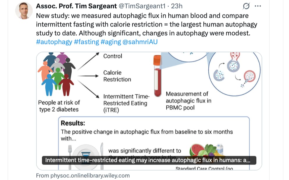
Tim Sergeant and his research team is moving forward on identifying the levels of autophagy activators; here comparing intermittent fasting vs. caloric restriction. I hope they do rapamycin soon too, so we have some basis of comparison.
Autophagy slows age-related pathologies and is stimulated by nutrient restriction in animal studies. However, this has never been shown in humans. We measured autophagy using a physiologically relevant measure of autophagic flux (flux of MAP1LC3B isoform II/LC3B-II in peripheral blood mononuclear cells in the context of whole blood) in 121 humans with obesity who were randomised to standard care (SC, control condition), calorie restriction (CR) or intermittent fasting plus time-restricted eating (iTRE) for 6 months. While the differences in change from baseline between groups was not significant at 2 months, we observed a significant difference in change from baseline between iTRE compared to SC at 6 months (P = 0.04, post hoc analysis). This effect may be driven partly by a tendency for autophagy to decrease in the SC group. The difference in change from baseline between CR and SC was not significant. Uncorrected analysis of correlations showed a negative relationship between change in autophagy and change in blood triglycerides. Data on the specificity and performance of the methods used to measure human autophagy are also presented. This shows autophagy may be increased by intermittent nutrient restriction in humans. If so, this is a demonstration that nutrient restriction can be used to improve a primary hallmark of biological ageing and provides a mechanism for how fasting could delay the onset of age-related disease.
Key points
- Autophagy slows biological ageing, and dysfunction of autophagy has been implicated in age-related disease – an effective way of increasing autophagy in cells and animal models is nutrient restriction.
- However, the impact of different types of nutrient restriction on physiological autophagic flux in humans has not been extensively researched.
- Here we measure the effect of intermittent time-restricted eating (iTRE) and calorie restriction on physiological autophagic flux in peripheral blood mononuclear cells.
- After 6 months, there was a significant difference in change from baseline between the iTRE and the standard care control group, with flux being higher in the iTRE group at this timepoint.
- However, there was no significant increase from baseline within the iTRE group, showing that although autophagy may be modified by nutrient restriction in humans, further studies are required.
https://physoc.onlinelibrary.wiley.com/doi/10.1113/JP287938
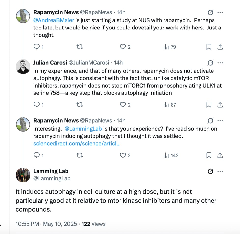
Interesting comment:
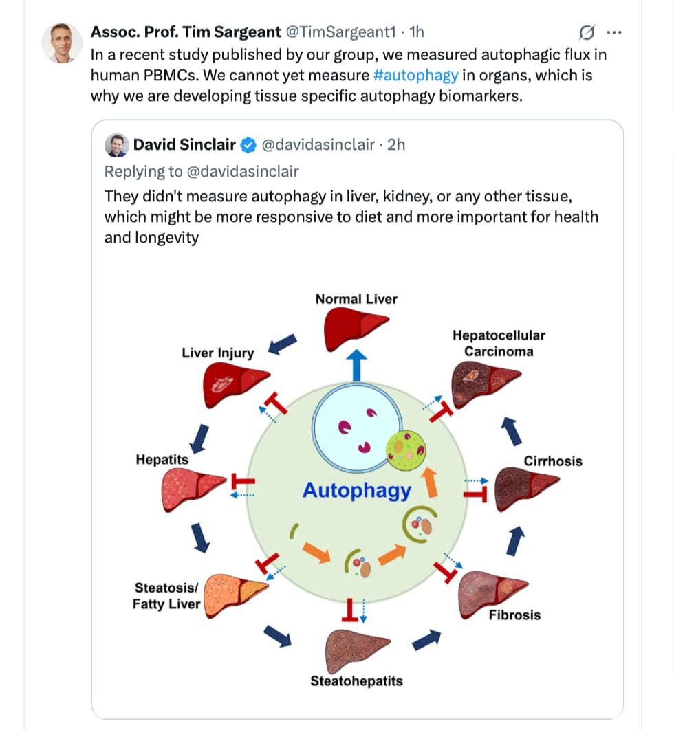
Source: https://x.com/TimSargeant1/status/1921007958579630401
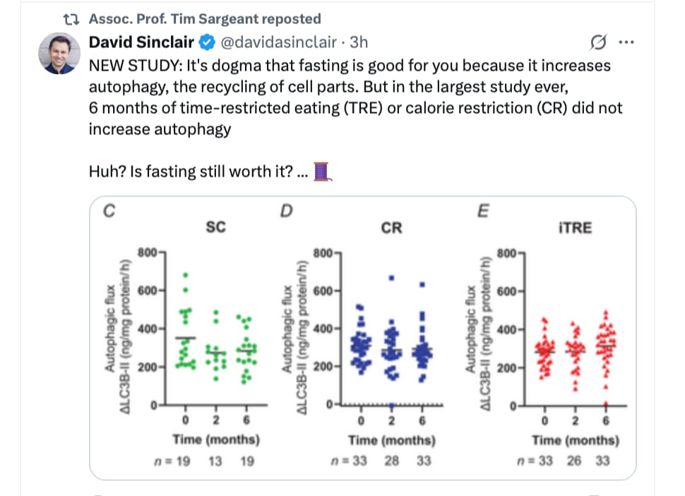
That’s very interesting and seems to challenge the fundamental belief or proof of Rapa inducing autophagy!
IF promoting autoaphagy as opposed to CR is not surprising.
May be best to continue to adhere to fasting on the first day of Rapa doing and then continue with IF.
IF in mice equals extended water fasting in humans. Not ideal for muscle preservation.