Cardiovascular Health 2026

OK, but did you know that the same thing applies to plant sterols :grin:? I would guess it’s logical. Undoubtedly it is a good thing in case of sitosterolemia. But is it also good in the absence of sitosterolemia, given that some have proposed - controversially - plant sterols as beneficial in CVD? Inquiring minds want to know. Any medication deserves a thorough establishment of all effects. A couple of papers (the EZE had a tiny # of subjects):

Ezetimibe Reduces Plant Sterol Accumulation and Favorably Increases Platelet Count in Sitosterolemia

A general look at phytosterols (review):

Phytosterols and Cardiovascular Disease

2 Likes

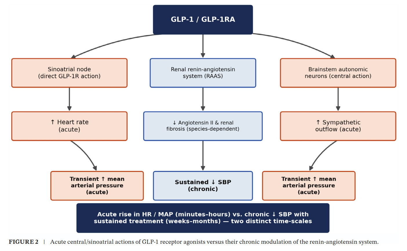
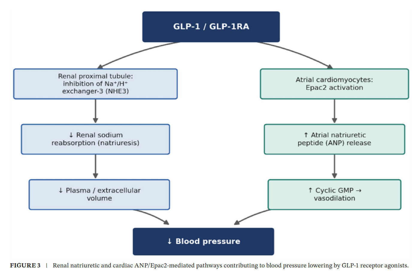
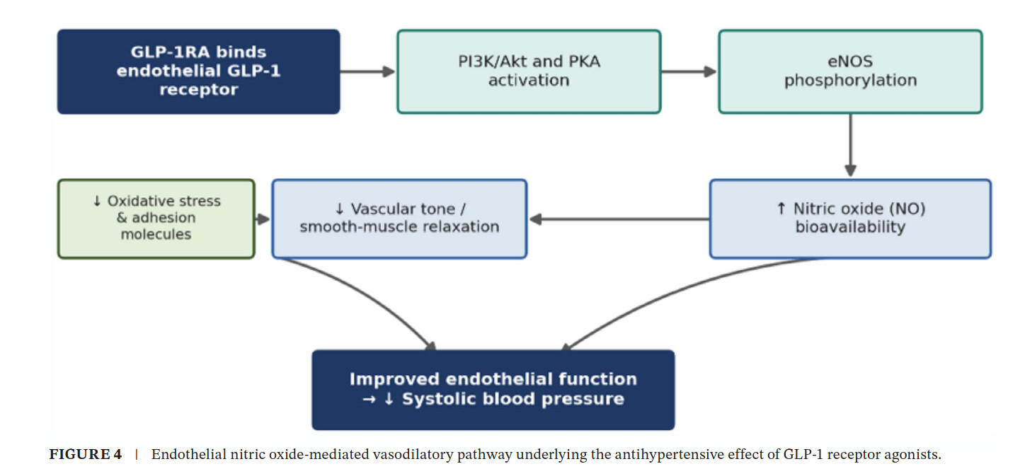
Glucagon-Like Peptide-1 Agonists and Blood Pressure Regulation: Molecular Mechanisms and Cardiovascular Implications

https://bpspubs.onlinelibrary.wiley.com/doi/epdf/10.1002/prp2.70329

AI summary:

1 Like

Let us keep in mind that rapamycin appears to polarize M1/M2 in the opposite direction from GLP-1RA. And in a contest between the two, rapamycin takes precedence as it’s upstream of where GLP-1RA work here and affects the very mechanism of polarization leaving GLP-1RA nothing to work with. If you are taking both, be aware that it is rapamycin that will be dominant. In this context pulsed administration of rapamycin becomes interesting, as one could speculate that in the trough of zero, GLP-1RA could step in to again re-balance M1 and M2.

However, the interplay of GLP-1 and rapamycin - for that matter other anti-diabetics too, is quite complex and frequently counter-intuitive. When I research a drug for inclusion into my stack, I spend a huge amount of time and effort trying to map out the possible interactions, because that is really where the rubber hits the road. It makes little sense to look at the actions of a drug on paper - such as the quote above “promotes M1 to M2 macrophage polarization, reduces pro-inflammatory cytokines” - if the other drugs in your stack completely abolish that effect, because you are counting on an effect that will be absent. You must then decide how to balance out these drugs by whatever means - dose, protocol, timing, or even inclusion.

Rapamycin and anti-diabetic drugs including GLP-1RA have a complex interplay in glucose control too. I have frequently noted in many threads that rapamycin definitely can be deleterious to pancreatic beta cells. This is a serious issue I have spent a lot of time on as someone who both takes rapamycin and has been pre-diabetic (for more than a decade).

Below is a paper that is super helpful in illuminating some of these issues, including the rapamycin and GLP-1 interplay, M1/M2 and other issues discussed. I didn’t post it before as it doesn’t fit neatly into any of the threads - rapamycin, glucose control, GLP-1 and anti-diabetics, pancreatic health. But because it focuses so much on the inflammatory aspect, I guess I’ll post it in this thread, even though it doesn’t really fit perfectly. I would strongly urge anyone interested in understanding these issues to read the whole paper to really get a good grounding in the underlying physiology. Enjoy!

mTOR: A double-edged sword for diabetes

https://jlb.onlinelibrary.wiley.com/doi/full/10.1002/JLB.3MR0317-095RR

5 Likes

Thank you for sharing—this is a very interesting paper.

The effect of rapamycin on β-cells and insulin resistance is a topic I’m particularly interested in. It provides insights that complement other papers I have read regarding its impact on calcium uptake.

I asked Opus 5.5 (extra high mode) to reconcile these papers, specifically focusing on my context: glucose dysregulation, pulsed rather than chronic rapamycin, and non-T1DM.

Here it is:

Short answer: the review doesn’t contradict the calcium idea, but it doesn’t support it either. It is about how diabetes develops (immune cells and islet growth) and rests almost entirely on rodent and cell-culture work. The calcium paper you’re remembering (Lombardi 2017) is real. Its “calcium” finding is best read as one side of a mitochondrial defect in how β-cells link glucose metabolism to insulin release. A second group described the same defect from the Krebs-cycle side. The bigger issue for you is different: nothing yet shows that a β-cell mechanism causes your ~1-week CGM rise. And your protocol has a built-in confound that could produce the same signal without rapamycin. I also need to retract several things I told you in May.

1. Which parts of the review apply to you

Review section Relevance Why
Immune half (NK, CD8, Th1/Th17, M1 cells destroying islets) None This describes autoimmune or inflammatory β-cell injury. Your GAD65, IA-2 and ZnT8 antibodies were all negative (2026-07-22) and hs-CRP is 0.44. Your NK count of 490 is a blood finding, not islet infiltration.
β-cell growth and mass (mTORC1 → cyclin D2) Negligible This is slow mass adaptation in rodents. It can’t produce a reversible 1-week signal.
mTORC2 → β-cell toxicity (Barlow 2012, ref 122) Possible, but needs prolonged exposure Rapamycin inhibited mTORC1 acutely, but the toxicity tracked mTORC2 dissociation and loss of PKB (Akt) activity. Knocking down RICTOR to inactivate mTORC2 selectively reproduced the toxicity of chronic rapamycin. (C)
Liver glucose production (Houde 2010, ref 53) A real candidate In rats given 2 mg/kg/day for 15 days, the liver switched on its glucose-production (gluconeogenic) program and caused severe glucose intolerance, even though hepatic insulin signalling to Akt was preserved. Chronic dosing again, and apparently not via mTORC2. (C)

The review also states that inhibiting mTOR promotes insulin resistance. That is the chronic picture. The one acute human study points the other way. With amino acids infused to switch on mTOR, rapamycin raised insulin-stimulated glucose uptake by 17% at meal-like insulin levels, with no effect at low insulin. (B: one clamp study in healthy volunteers.) You eat 1.6 g/kg of protein and take leucine during runs. An acute pulse is therefore more likely to help muscle insulin action than hurt it. Peripheral insulin resistance is the least likely acute driver for you.

2. What the calcium paper actually showed

Lombardi 2017 tested rat β-cell lines plus mouse and human islets at 25 nM for 24 h:

  • Glucose-stimulated insulin secretion fell; viability was unchanged; secretion in response to leucine and glutamine was impaired while the response to KCl (direct depolarisation) was intact; oxygen consumption fell; ER Ca²⁺ stores were depleted with increased leak; mitochondrial Ca²⁺ uptake fell; and all three IP3R isoforms were upregulated, which the authors proposed as the mechanism.
  • The intact KCl response matters. The insulin-release machinery itself works; the lesion sits upstream, in metabolism.

Shimodahira 2010 (cited in the review) used rat islets for 24 h:

  • ATP fell, glucose oxidation fell, and α-ketoglutarate dehydrogenase (α-KGDH) activity fell, while the respiratory chain downstream of complex II was unaffected.

These two findings likely describe one lesion. α-KGDH is one of the mitochondrial enzymes that calcium switches on. So less mitochondrial Ca²⁺ uptake (Lombardi) and lower α-KGDH activity (Shimodahira) are plausibly the same defect seen from opposite ends. Calcium is a node in that loop, not necessarily its root. (C: this link is my synthesis; neither paper tested it.)

What the paper does not show:

  • Which mTOR complex is involved. It was never tested. The closest data (Barlow, also in human islets) point to mTORC2 under prolonged exposure.
  • How long the effect lasts. There was no washout experiment, so nothing tells us about recovery after the drug clears.
  • Whether the dose matches yours. The 25 nM figure comes from Desai 2003, which measured portal-vein drug levels in islet-transplant recipients. That is continuous dosing, sampled where the islets were infused.
    • The label gives a blood-to-plasma ratio of 36 ± 18, meaning the drug sits mostly in blood cells, with about 92% of plasma drug bound to protein.
    • So free drug ≈ whole blood ÷ 36 × 0.08 ≈ 0.2% of the whole-blood level. A whole-blood 23 ng/mL (= 25 nM) works out to about 0.05 nM free.
    • Culture medium has no red cells and far less protein, so most of the 25 nM in a dish reaches the cells.
    • Caveats: red-cell binding saturates, so the free fraction at your peak is higher than this. Rapamycin also accumulates inside cells bound to FKBP12.
    • Net: the cell-culture magnitude is an upper bound for your pulse, not an estimate. (C)

Sources:

1 Like

Calcium handling here is a huge area, and would take us way outside the remit of the subject matter of this thread, so I’m not going to comment on it, other than to signal yet again that in my view the impact of rapamycin on pancreatic beta cells is complex and overall deleterious. However… it is complex, meaning you can find rapamycin on both sides of the issue (especially when you look at endoplasmic reticulum stress in beta cells). And because when it comes to discussing medical issues I have impulse control problems, I cannot restrain myself from posting some important papers (apologies - it has nothing to do directly with “cardiovascular health 2026”).

Glucose Amplifies Fatty Acid-Induced Endoplasmic Reticulum Stress in Pancreatic β-Cells via Activation of mTORC1

Autophagy plays a protective role in endoplasmic reticulum stress-mediated pancreatic β cell death

https://www.tandfonline.com/doi/full/10.4161/auto.21994

It is not for no reason that rapamycin coupled with glucose control agents results in superior health/longevity outcomes. Specifically, I believe it has to do with those agents which lower insulin demand, release and overall levels. Acarbose for example prevents the sharp bolus demand for insulin by eliminating glucose spikes. That leads to lower insulin demand and doesn’t hammer beta cells as much. It is especially important to take an SGLT2i if you are on rapamycin, because you are dumping glucose which doesn’t need to be handled by more insulin release. When taking rapamycin, you should adopt all measures to lessen stress on pancreatic beta cells.

3 Likes

:warning:CAUTION: Chinese paper :warning:

Lactoferrin influences atherosclerotic progression by modulating macrophagic AMPK/mTOR signaling-dependent autophagy

“This study aimed to explore the role of lactoferrin (LTF) in atherosclerosis (AS) and its possible mechanisms. Human left coronary artery tissues were collected and divided into control (CON), coronary heart disease (CHD) and sudden coronary death (SCD) groups. Pathologic changes (including changes in the coronary plaque area, necrotic core, collagen fibers, and foam cell content) were observed. The LTF, P62, and 4-hydroxynonenal (4-HNE) expression levels were assessed. The ApoE –/– AS mouse model was established. The pathological changes and related protein levels were analyzed after autophagy inhibition. The foam cell model was constructed using an ox-LDL-induced human monocyte line, THP-1. The LTF, BECN1, LC3-II/I, AMP-activated protein kinase (AMPK)/the mammalian target of rapamycin (mTOR) pathway proteins, B-cell lymphoma-2 (Bcl-2), Bcl-2-associated X protein (Bax), and 4-HNE expressions were then detected after silencing of LTF or BECN1. Plaque stability was significantly lower in the SCD group compared to the non-SCD group (p < 0.05). LTF, P62 and 4-HNE levels in plaques increased as plaque stability decreased, and LTF was significantly correlated with plaque progression and autophagy levels. Autophagy inhibition by U0126 leads to the worsening of aortic luminal stenosis, increased necrotic core and foam cell deposits, decreased autophagosomes, reduced LTF expression, and upregulated P62 expression in AS mice. It was further demonstrated that LTF expression correlates with autophagy. LTF expression was increased in ox-LDL-treated THP-1 cells, and silencing BECN1 and/or LTF increased mTOR phosphorylation and 4-HNE levels, inhibited BECN1 and LC3 II expression and AMPK activation, and simultaneously decreased the Bcl-2/Bax ratio. LTF might alleviate AS pathology through accelerating the AMPK/mTOR pathway, and suggested that LTF may be a potential predictive molecule for AS.”

1 Like

How does ezetimibe impact fatty acid absorption?

Regarding ALA (alpha linolenic acid - short chain n3 poly found in vegetarian sources like flaxseed and walnuts): ezetimibe strongly inhibits absorption… or maybe not. All in cardiac disease patients.

A randomized trial of the effects of ezetimibe on the absorption of omega-3 fatty acids in cardiac disease patients: A pilot study

https://www.sciencedirect.com/science/article/abs/pii/S2405457715001102

“Elevated levels of circulating omega-3 polyunsaturated fatty acids like alpha linolenic acid (ALA) may be beneficial for cardiovascular health. Circulating ALA concentrations are elevated dramatically by a cholesterol supplemented diet which increases ALA bioavailability through enhanced micelle formation in the intestines.”

“Ezetimibe therapy inhibited the absorption of omega-3 fatty acids. Patients receiving ezetimibe therapy may not receive the expected cardiovascular benefits from dietary supplementation with omega-3 fatty acids.”

However, another (weaker) study claims ezetimibe does not affect the absorption of any PUFA, long or short chain EPA, DHA, n3, n6. Possibly mediated by concurrent use of high vs low dose statin.

Effects of Ezetimibe on Serum Polyunsaturated Fatty Acids in Patients With Coronary Artery Disease

https://www.jstage.jst.go.jp/article/ihj/54/5/54_254/_pdf/-char/en

“In conclusion, ezetimibe in combination with a low-dose statin, or as monotherapy in statin-intolerant patients, decreased LDL cholesterol, but did not significantly affect serum PUFA concentrations in patients with coronary artery disease.”

IN MICE ezetimibe inhibited absorption of saturated fatty acids.

Reduced absorption of saturated fatty acids and resistance to diet-induced obesity and diabetes by ezetimibe-treated and Npc1l1 −/− mice

And I’m putting this here as the idiotic software does not allow me to make more than three posts without a reply from someone (moronic restriction):

A systematic review and meta-analysis of randomized controlled trials on the impact of ezetimibe on carotid intima-media thickness

https://www.jns-journal.com/article/S0022-510X(23)02090-7/fulltext

“In elderly individuals, non-familial hypercholesterolemia, and secondary prevention, the impact of ezetimibe on CIMT decrease was demonstrated. These findings imply that ezetimibe can reduce the incidence and recurrence of stroke.”

Here ya go!!!

2 Likes

Both T2DM and CVD discussed in the paper.

Peanuts-derived plasma lipids and lower risk of type 2 diabetes: association mediated by body mass index—an intervention and case-cohort study

https://www.sciencedirect.com/science/article/pii/S0002916526003369?via%3Dihub

Pop-sci article:

Peanut consumption affects the lipid profile circulating in the blood

https://medicalxpress.com/news/2026-10-peanut-consumption-affects-lipid-profile.html

“New findings from an intervention and cohort study suggest that the fatty acids provided by peanuts do more than just contribute calories. Researchers at the German Institute of Human Nutrition Potsdam-Rehbruecke (DIfE), together with colleagues at the University of Barcelona, have demonstrated that a specific “Peanut Lipidomic Signature” (PeLS) is associated with a reduction in the risk of type 2 diabetes. The study was published in The American Journal of Clinical Nutrition , and reveals how peanut consumption triggers a measurable remodeling of the human lipidome.“

2 Likes

Intensive vs Standard Blood Pressure Lowering in People With and Without Chronic Kidney DiseaseA Systematic Review and Meta-Analysis

https://jamanetwork.com/journals/jamacardiology/fullarticle/2854798

" Key Points

Question What are the effects of intensive blood pressure (BP) lowering on cardiovascular and kidney outcomes among adults with and without chronic kidney disease (CKD)?

Findings This systematic review and meta-analysis indicates that in people with and without CKD, intensive BP lowering reduced risk of cardiovascular events by 15% per 10-mm Hg lower systolic BP. Intensive BP lowering did not affect risk of kidney failure but increased risk of sustained declines in kidney function.

Meaning Intensive BP lowering should be recommended in people with and without CKD to reduce cardiovascular risk; clinicians should implement BP-lowering strategies with careful monitoring of kidney function, despite no increased risk of kidney failure.

Abstract

Importance There is uncertainty about the effects of intensive blood pressure (BP) lowering on risks of cardiovascular disease and kidney failure in patients with chronic kidney disease (CKD).

Objective To assess the standardized effects of intensive BP lowering on cardiovascular and kidney outcomes overall, and then to assess for effect modification by CKD status.

Data Sources MEDLINE and Embase databases were searched from inception to January 16, 2026.

Study Selection Randomized clinical trials comparing intensive vs standard BP lowering in adults with and without CKD, with at least 1 year of follow-up, were eligible for inclusion.

Data Extraction and Synthesis Meta-analyses were performed using inverse variance weighting after standardizing trial results according to the between-group difference in achieved systolic BP (SBP). Differences in relative effects between CKD and non-CKD populations were assessed using χ2 tests for heterogeneity.

Main Outcomes and Measures The primary outcome was major cardiovascular events; secondary outcomes included kidney failure and estimated glomerular filtration rate (eGFR)–based surrogate outcomes for kidney failure.

Results We identified 27 studies including 88 830 participants. Intensive BP lowering reduced the risk of cardiovascular events by 15% per 10-mm Hg lower SBP (relative risk [RR], 0.85; 95% CI, 0.81-0.89), without heterogeneity by CKD status. This corresponded to an estimated (SE) absolute reduction of 4.3 (1.2) cardiovascular events per 1000 person-years in individuals with CKD and 2.7 (0.4) events per 1000 person-years in those without CKD. Intensive BP lowering did not affect risk of kidney failure (RR per 10-mm Hg reduction, 0.97; 95% CI, 0.84-1.11) nor risk of 57% or 50% sustained eGFR declines (RR, 0.97; 95% CI, 0.83-1.14; and RR, 1.29; 95% CI, 0.94-1.76, respectively), without heterogeneity by CKD subgroup. Intensive BP lowering increased risk of 30% or greater and 40% or greater declines by 42% (RR, 1.42; 95% CI, 1.28-1.58) and 47% (RR, 1.47; 95% CI, 1.30-1.66).

Conclusions and Relevance Results of this systematic review and meta-analysis indicate that in adults with or without CKD, intensive BP lowering reduced the risk of cardiovascular events by 15% per 10-mm Hg lower SBP and increased the risk of sustained declines in eGFR, without an increase in risk of kidney failure. However, since baseline cardiovascular risk is higher in CKD, the absolute reduction in cardiovascular events was at least as large in this population; therefore, intensive BP lowering should be recommended in people with and without CKD."

1 Like

I had the impression that higher BP was damaging for kidneys, but it appears that lower BP makes them work less well even if functionally they are much the same.

It might be a short term effect, akin to SGLT2i also initially lowering GFR, before recovering within a year or so, except the SGLT2i really slow down the natural age related GFR decline over time; the lower BP might not have the same effect size. Perhaps also it might matter which meds were used to lower BP - diuretics, CCB, ACEi, ARB all might have differential effect on kidneys apart from BP lowering (incidentally SGLT2i lower BP very slightly in hypertensives).

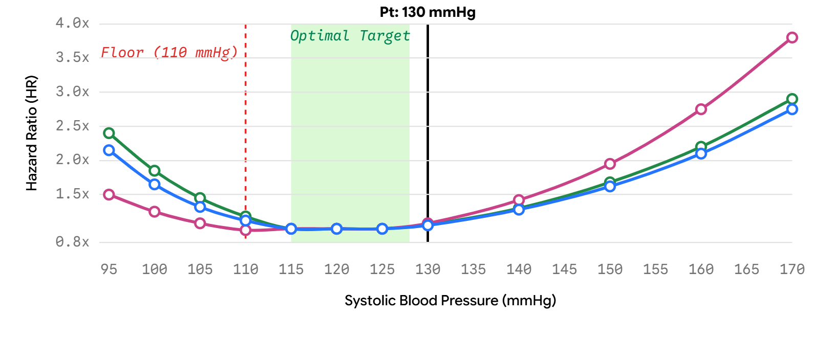
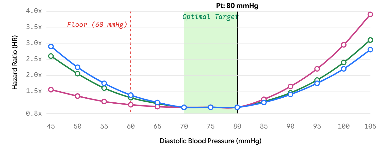
It appears that I have been overdosing on telmisartan. Any drug that lowers systolic also lowers diastolic. I did this by increasing my telmisartan dose from 40 mg to 80 mg daily, based on previous discussions about telmisartan. Another reminder to double- and triple-check the self-medication use based on user discussions. Also, the recommended systolic, and especially diastolic, blood pressure is a little higher than some discussions suggest.

The following is from Gemini Pro Argon and Aristotle:

Lowering blood pressure almost always causes an initial, measurable drop in estimated Glomerular Filtration Rate (eGFR). This happens because the kidneys rely on incoming vascular pressure to push blood through their microscopic filters (glomeruli).

There is a definitive threshold where lowering blood pressure transitions from protecting the kidneys to depriving them of necessary blood flow, a state known as renal hypoperfusion.

The Mechanics of eGFR and Blood Pressure

To understand how blood pressure alters eGFR, it helps to look at the mechanics of the kidney’s filtration system:

  • The Pressure Dependency: The kidneys maintain a precise internal pressure to filter waste. When systemic blood pressure drops, the driving force behind this filtration decreases, leading to a lower eGFR.
  • The Medication Effect: Antihypertensive medications—specifically ACE inhibitors (like lisinopril) and ARBs (like telmisartan or losartan)—work by relaxing the efferent arteriole, the exit vessel of the kidney filter. This intentionally lowers pressure inside the kidney to prevent long-term damage, but it predictably drops eGFR by 10% to 30% in the first few weeks of use. This initial dip is generally considered a sign that the drug is working to relieve stress on the kidneys, not a sign of kidney injury.
  • The Tipping Point: As we age, renal arteries often become stiffer or narrower. An 85-year-old vascular system may require a slightly higher systemic “driving pressure” to force blood into the kidneys compared to a younger person. If systolic blood pressure is pushed too low (often below 110 mm Hg in older adults, though it varies heavily by individual), the kidneys can no longer autoregulate. They become starved for oxygen and blood flow, causing eGFR to plummet abruptly and waste products to build up in the blood.

If blood pressure is being lowered and eGFR drops, utilizing a Cystatin C blood test provides a much clearer picture. Cystatin C is a protein produced by all cells at a steady rate and is completely unaffected by muscle mass, dietary protein, or recent gym sessions. Relying on a Cystatin C-based eGFR ensures that any observed drop in kidney function is truly a result of the blood pressure changes, rather than a harmless artifact of maintaining physical strength.

8 Likes

Source: This post on X

1 Like

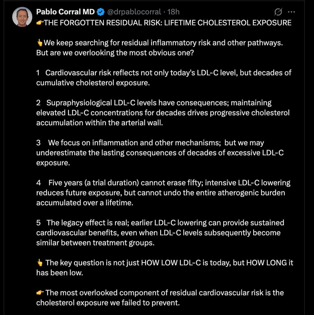
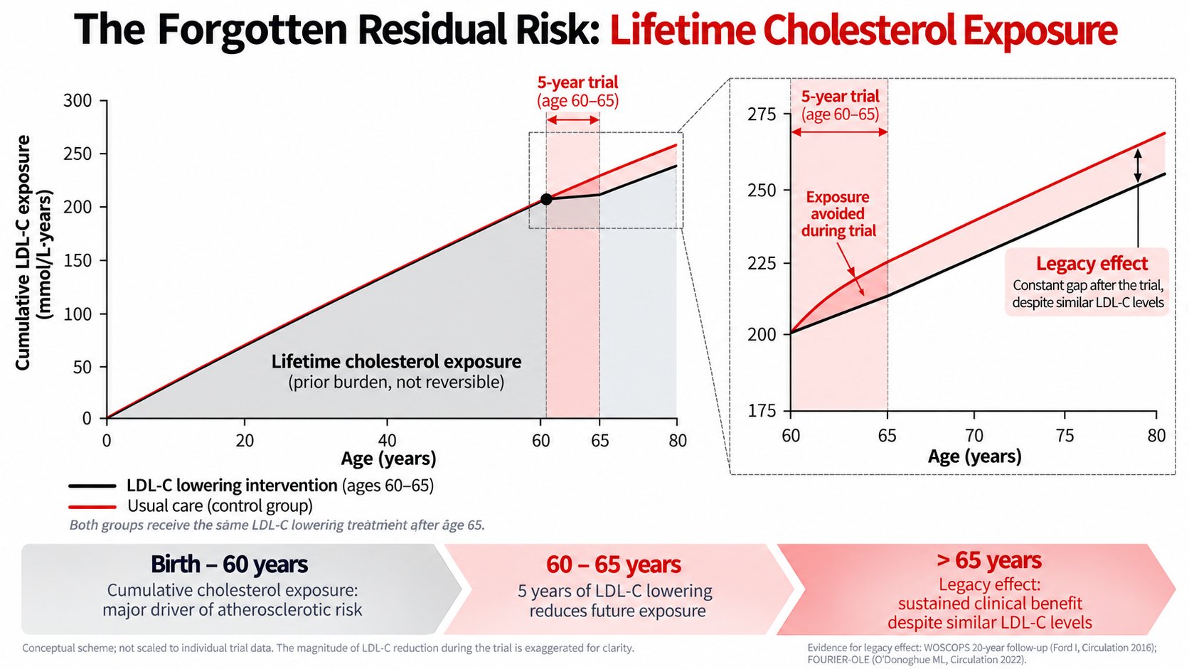
“The most overlooked” - OK, but that’s where imaging comes in. If you can see and estimate the amount of harm done by the amount of plaque present, then you are no longer “overlooking” the consequences of decades of high LDL exposure. That is why it’s an important tool - which we need to refine, as it’s central in how we look at atherosclerosis. Also, just assessing and knowing the residual risk doesn’t mean you necessarily have the tools to affect it. The damage is done. You have X amount of plaque - you can try to reverse the soft plaque burden, but there’s only one mechanism (if it even works): driving ApoB down to very low levels. And guess what - that’s what you should be doing regardless of what the imaging says. Therefore this residual risk is not actionable - you do what you’d have done regardless of how much of this residual risk (plaque) you have or don’t have - if “have” drive ApoB low, if “don’t have” drive ApoB low to keep it that way, and that’s that.

2 Likes

As you most likely have seen, I occasionally vacillate on getting a CTTA with CLEERLY. I finally messaged my past cardiologist Dr Yeghiazarians at UCSF (he’s the best one I’ve had but he booted me out of his practice :slight_smile: ) and asked him…

Regarding all the various imaging options, he said:

“none of them will change the recommendations from a clinical management standpoint as stated above assuming you are asymptomatic from cardiovascular standpoint.”

Aka …don’t waste your time and money (for me)

This doesn’t mean I don’t continue to be super curious…and I will most likely continue to vacillate

4 Likes

This is exactly my line of thinking. If I have no symptoms of any CVD whatsoever and If I’m doing everything right to protect my heart (against CVD) then my thinking is that I MUST continue to be symptom free, which means CVD free. Why bother or why spend considerable amount of money to find out either way when in fact even if it showed I have plaque it would not change a thing in my stack as I’m already doing everything right (i.e. Ezetimibe, PITA, Fish oil, no unhealthy fats in the diet, plenty of veggies, exercise etc.). So, I decided to save the money and continue what I’m doing, and I may never know if I have any plaque LOL.

3 Likes
4 Likes

Good to see some of these blood pressure studies… I hope, the lower the better…

Since being on solid dosed reta for about a year my fat is at zero %… And with my lengthy supplement stack including novos, and all my prescriptions that can lower BP as well - including jardiance and Telmisartan 80 and high dose statins, my BP can sit near hypotension 90/60, particularly when its at the lowest point of the day which is when I wake up for me…

I made efforts to try to increase the BP, even getting off Telmisartan for a time, but in the end, just left it there as the blood work never was impeded by the low BP

I do have mild side effects, like if I jump up after laying down I can get dizzy (but I do not have orthostatic hypotension as measured by my doctor in the hospital)… but I’m still kind of just hoping, like LDL, if your seriously asymptomatic, the lower the better, lol (if a disease/lack of healthy isnt causing the low BP). After all, its healthy substances which are creating the near hypotension

1 Like

Just be careful. When my blood pressure got too low, I passed out in the toilet and hit my head. It could have killed me, but I was lucky.

The tell for passing out was I felt nauseous which is why I headed to the bathroom.

8 Likes